
Asistență medicală

Online, de la tine de acasă!

Personal profesionist

Doar doctori oncologi de top

Încărcați dosarul cazului dvs.

Și îl vom cerceta!

Blog

Endometrial cancer

Endometrial cancer

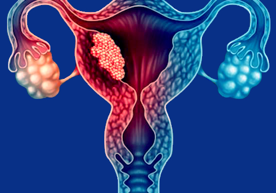
Endometrial cancer is a type of cancer that begins in the uterus. The uterus is the hollow, pear-shaped pelvic organ where fetal development occurs.

Endometrial cancer begins in the layer of cells that form the lining (endometrium) of the uterus. Endometrial cancer is sometimes called uterine cancer. Other types of cancer can form in the uterus, including uterine sarcoma, but they are much less common than endometrial cancer.

Endometrial cancer is often detected at an early stage because it frequently produces abnormal vaginal bleeding. If endometrial cancer is discovered early, removing the uterus surgically often cures endometrial cancer.

Symptoms

Signs and symptoms of endometrial cancer may include:

  • Vaginal bleeding after menopause
  • Bleeding between periods
  • Pelvic pain

When to see a doctor

Make an appointment with your doctor if you experience any persistent signs or symptoms that worry you.

Causes

Doctors don’t know what causes endometrial cancer. What’s known is that something occurs to create changes (mutations) in the DNA of cells in the endometrium — the lining of the uterus.

The mutation turns normal, healthy cells into abnormal cells. Healthy cells grow and multiply at a set rate, eventually dying at a set time. Abnormal cells grow and multiply out of control, and they don’t die at a set time. The accumulating abnormal cells form a mass (tumor). Cancer cells invade nearby tissues and can separate from an initial tumor to spread elsewhere in the body (metastasize)

Risk factors

Factors that increase the risk of endometrial cancer include:

  • Changes in the balance of female hormones in the body. The ovaries make two main female hormones — estrogen and progesterone. Fluctuations in the balance of these hormones cause changes in the endometrium.A disease or condition that increases the amount of estrogen, but not the level of progesterone, in your body can increase your risk of endometrial cancer. Examples include irregular ovulation patterns, which might happen in polycystic ovary syndrome, obesity and diabetes. Taking hormones after menopause that contain estrogen but not progesterone increases the risk of endometrial cancer.A rare type of ovarian tumor that secretes estrogen also can increase the risk of endometrial cancer.
  • More years of menstruation. Starting menstruation at an early age — before age 12 — or beginning menopause later increases the risk of endometrial cancer. The more periods you’ve had, the more exposure your endometrium has had to estrogen.
  • Never having been pregnant. If you’ve never been pregnant, you have a higher risk of endometrial cancer than someone who has had at least one pregnancy.
  • Older age. As you get older, your risk of endometrial cancer increases. Endometrial cancer occurs most often after menopause.
  • Obesity. Being obese increases your risk of endometrial cancer. This may occur because excess body fat alters your body’s balance of hormones.
  • Hormone therapy for breast cancer. Taking the hormone therapy drug tamoxifen for breast cancer can increase the risk of developing endometrial cancer. If you’re taking tamoxifen, discuss this risk with your doctor. For most, the benefits of tamoxifen outweigh the small risk of endometrial cancer.
  • An inherited colon cancer syndrome. Lynch syndrome, also called hereditary nonpolyposis colorectal cancer (HNPCC), is a syndrome that increases the risk of colon cancer and other cancers, including endometrial cancer. Lynch syndrome is caused by a gene mutation passed from parents to children. If a family member has been diagnosed with Lynch syndrome, discuss your risk of the genetic syndrome with your doctor. If you’ve been diagnosed with Lynch syndrome, ask your doctor what cancer screening tests you should undergo.

Prevention

To reduce your risk of endometrial cancer, you may wish to:

  • Talk to your doctor about the risks of hormone therapy after menopause. If you’re considering hormone replacement therapy to help control menopause symptoms, talk to your doctor about the risks and benefits. Unless you’ve undergone a hysterectomy, replacing estrogen alone after menopause may increase your risk of endometrial cancer. Taking a combination of estrogen and progestin can reduce this risk. Hormone therapy carries other risks, so weigh the benefits and risks with your doctor.
  • Consider taking birth control pills. Using oral contraceptives for at least one year may reduce endometrial cancer risk. The risk reduction is thought to last for several years after you stop taking oral contraceptives. Oral contraceptives have side effects, though, so discuss the benefits and risks with your doctor.
  • Maintain a healthy weight. Obesity increases the risk of endometrial cancer, so work to achieve and maintain a healthy weight. If you need to lose weight, increase your physical activity and reduce the number of calories you eat each day.


Can Radiation Be Given Twice in the Same Area?

Healthy cells in the human body divide and grow. Cancer cells do this at a much faster rate. Radiation therapy, or radiotherapy, is a cancer treatment that uses high-energy beams to damage or destroy fast-growing cancer cells. Radiation therapy breaks the DNA of cancer cells and prevents them from growing. While chemotherapy and certain other cancer treatments are systemic (they expose the entire body to the drug), radiation therapy is a local treatment. It is aimed at only the part of the body affected by cancer. Nonetheless, some normal cells nearby are affected by the radiation treatment, although most recover function. 

More than half of all people with cancer get some form of radiation treatment. Sometimes radiation is the only treatment required. At other times, it is given along with other types of therapies to treat cancer, such as chemotherapy and surgery. 

Radiation treatments have been used for more than 100 years now. Medical advances during this time have made radiation therapy safe and effective. Radiation oncologists can plan the radiotherapy so that it damages cancer cells while causing as little harm as possible to healthy cells. Throughout the treatment, patients are monitored to check how the cancer is responding. 

Please continue reading to learn more about radiation therapy protocols, the dose of radiation, side effects of radiation, and whether cancer patients can get radiotherapy twice in the same area.

What are the different types of radiation therapy?

External beam radiation therapy

This is a type of radiation treatment in which a beam of high-energy X-rays is directed at the cancer through the skin. The radiation oncology team controls the size and shape of the beam and its direction on the body to treat the tumor while sparing surrounding healthy tissue. Different types of external beam therapy are used to treat specific types of cancers.

Three-Dimensional Conformal Radiation Therapy (3D-CRT)

3D-CRT uses computer imaging to map the size, shape, and location of a tumor (tumors come in various shapes and sizes and are not regular). CT, MRI, and PET scanning create detailed 3D representations of the tumor and the surrounding structures. This allows the radiation oncologist to precisely target the tumor with the radiation beam with very little damage to nearby normal tissue, allowing them to heal quickly.

Intensity Modulated Radiation Therapy (IMRT)

IMRT is a special type of 3D-CRT that allows the radiation beam to match the tumor’s shape more precisely. In IMRT, the beam is broken into smaller “beamlets.” Each beamlet can be adjusted individually for intensity. This allows the radiation oncologist to limit damage to healthy surrounding tissue further. IMRT, in some cases, may allow a higher radiation dose to be delivered to the tumor, possibly increasing the chance of a cure.

Proton Beam Therapy and Neutron Beam Therapy

These are specialized radiation therapy techniques that use protons and neutrons instead of X-ray to treat cancer. The physical characteristics of these high-energy particles allow doctors to reduce the radiation doses to surrounding healthy tissue. These radiation beams also have a greater biological impact on tumors that can be advantageous in treating certain cancers. Unfortunately, this type of radiation treatment is available only at a few specialized radiation oncology centers.

Stereotactic Body Radiation Therapy

Stereotactic body radiotherapy is a radiation treatment technique that allows doctors to focus radiation beams on certain tumors more precisely. The additional precision is achieved by using immobilization, such as a head frame to treat head and neck cancers. This type of radiation may be given as a single dose and is sometimes called radiosurgery. It may also be given as 3-8 treatments for a tumor outside the brain. 

Image-Guided Radiation Therapy (IGRT)

Brachytherapy

This type of “internal radiation” involves placing radioactive material into or near a tumor. The placement of the radiation source close to the tumor allows the radiation oncologist to directly deliver a large dose of radiation to the cancer cells. The radioactive sources may be left in place permanently or temporarily to treat cancer. 

There are other types of radiation therapy besides the common ones mentioned above.

Can radiotherapy be repeated?

IGRT helps deliver radiation to a tumor more precisely by using imaging (CT, ultrasound, X-rays). Images are obtained just before the radiation therapy is given and compared to earlier images to see if the radiation treatment needs to be adjusted. 

The goal of any cancer treatment is to control the growth of the tumor with minimal side effects and minimal impairments in the patient’s quality of life. In the past, patients who received radiation therapy once could not be treated with radiation a second time. This was because the area targeted by the radiation could not be mapped precisely enough. As a result, the healthy tissue surrounding the tumor was at risk of a radiation overdose. Survival rates after repeat radiation treatment were not good. Life-threatening side effects meant that only 50% of all patients who received repeat radiation therapy were alive after one year, and only 20% were alive after three years. Also, there is a long-term risk of developing a second cancer from the radiotherapy itself. While this risk is low, it depends on how much radiation is given. A second round of radiation treatment can increase the risk of a second cancer.

The body doesn’t forget its first encounter with radiation, so you can’t treat it a second time like you did the first time. 

In recent years, however, advances in radiation oncology have made it possible for some patients to undergo repeat radiotherapy. In particular, repeat external beam radiotherapy for locally recurrent head and neck tumors has been given with an intent to cure. Unfortunately, this is possible in only a few carefully selected patients. The initial results from a few small trials are encouraging, but repeat radiation therapy remains experimental. 

To undergo repeat radiotherapy, patients are selected by an interdisciplinary tumor board. Repeat radiation therapy is usually carried out in a specialized radiation oncology center. 

How many times can you have radiation therapy?

As noted above, repeat radiation therapy has been tried in some types of head and neck cancers. However, in most patients, radiation treatment is given once. To qualify as a potential candidate for the second round of radiotherapy, the patient must meet certain conditions, including a good general health status, a small localized recurrence of the tumor that is well-circumscribed, clean surgical margins, more than 6 months having passed since the initial round of radiation therapy, availability of documentation on the initial radiotherapy for evaluation, a reserve capacity in the surrounding normal tissue to withstand radiation, and assessment and recommendation by an interdisciplinary cancer treatment team.

Notably, a second round of radiation therapy alone may not work. Without surgery, cancer has a high chance of coming back. A crucial part of successful treatment of the disease is a collaborative approach with the radiation oncologist working with the surgeon. 

Can you have radiation twice in one day?

In general, the answer is no. Patients do not receive radiation twice in one day. However, radiation oncologists have experimented with giving a higher dose of radiation with each treatment to try and shorten the course of radiation treatment (for example, to get a 6-week radiation treatment down to 5 weeks or 4 weeks). Clinical studies have shown encouraging results in this regard. If the results are found to sustain after a longer period of evaluation (patients remain cancer-free), then in the future, we may see courses of radiation becoming shorter by giving a little more radiation dose with each treatment, but not by coming in more than once a day.

How long do you have to wait between radiation treatments?

The usual protocol for external beam radiation therapy is 5 treatment sessions per week, Monday through Friday, for 3-9 weeks, depending on the type of cancer and specific treatment plan. The 2-day break each week allows the body to repair some of the damage caused by the radiation to healthy tissues. However, some side effects may not go away until the radiation treatment is completed. 

If you are deemed an appropriate candidate for a second round of radiation therapy, your radiation oncologist will likely advise waiting at least 6 months after the first course of radiotherapy.

A new beginning, from us for you 🎗️ 🎗️ 🎗️

According to the World Health Organisation, cancer is the second leading cause of mortality worldwide, with roughly 9.6 million deaths due to one or more forms of the disease. In your opinion, how would you describe the current state of cancer drug discovery?

In the last few decades, we have made significant strides, but there is much more we can do: find more targeted treatments with less impactful side effects and address many of the cancers of unmet need, such as brain tumors and liver cancers.

We still do not understand these cancers well, so there must be an increased focus on investing in and collaborating on these areas across the industry – the more we know about these diseases, the better we can target them, and the kinder the treatments will be for patients.

We have seen massive steps taken to accelerate drug discovery. For example, the whole era of functional genomics and the leaps in the application of machine learning to aid disease targeting and wider drug discovery have helped us understand the causes, consequences and potential treatments of various cancers.

What are the major challenges that this cancer research faces?

Public spending and the general cost-of-living represent massive challenges for the charity sector and drug discovery projects funded by donations. When people’s budgets are limited, donating is understandably not always the first priority. In these times, we need to be mindful and try to mitigate the challenge to ensure our science does not suffer.

Another key area we need to improve on is early disease diagnosis. As with anything, early diagnosis provides the best chance for treatment or management. There is so much more we need to do to balance out what opportunities people have for early therapies as well as the range of treatments available to them.

However, this cannot be pursued in isolation. Early diagnosis must also consider and combat the huge inequalities existing globally across both diagnostic and cancer treatments, including those arising from ethnicity and socio-economic deprivation. CRUK and others are investing heavily to address this.

Combination of temozolomide and capecitabine associated with improved pfs compared to temozolomide alone in advanced pancreatic nets

A randomised, multicentre, phase II ECOG-ACRIN E2211 study of patients with advanced progressive pancreatic neuroendocrine tumours (NETs) met primary endpoint as treatment with temozolomide and capecitabine resulted in a prolonged median progression-free survival (PFS) compared to temozolomide alone (22.7 months versus 14.4 months). This clinically and statistically significant improvement in PFS with temozolomide and capecitabine is encouraging when analyzing the treatment landscape for patients with progressive pancreatic NETs. The response rate (RR) of 39.7% with temozolomide and capecitabine is higher than with other available treatments. In addition, the study investigators found that methylguanine methyltransferase (MGMT) deficiency is associated with tumour response. The study findings are reported by Dr. Pamela L. Kunz of the Yale Cancer Center in New Haven, CT, US and study colleagues on 19 October 2022 in the Journal of Clinical Oncology.

The authors reported in the background that patients with advanced pancreatic NETs have few treatment options that yield objective radiographic tumour regression. Prospective studies evaluating everolimus and sunitinib in this patient population have demonstrated prolonged PFS compared to placebo; however, RRs with these agents are less than 10%. 177Lu-DOTATATE, a novel radiopeptide, is approved for use in gastroenteropancreatic NETs based on both, the randomised NETTER-1 study in midgut NETs and retrospective studies in pancreatic NETs. However, it has not been formally evaluated in prospective randomised clinical studies in patients with pancreatic NETs, therefore limiting an accurate estimate of PFS.

Recent retrospective series and small, prospective phase II studies suggest that temozolomide is similarly active, but less toxic than streptozocin-based treatment in patients with pancreatic NETs. Additionally, temozolomide has been investigated prospectively in patients with NETs in small phase II combination studies. Studies evaluating single-agent capecitabine are limited to retrospective or small prospective studies and show little to modest activity. However, pre-clinical and early clinical evidence suggest that capecitabine may be synergistic with temozolomide, perhaps by downregulating the DNA-repair enzyme, MGMT.

Furthermore, temozolomide, like streptozocin, is an alkylating agent that induces DNA methylation at the O6 position of guanine leading to DNA damage and cell death, usually repaired by MGMT. In glioblastoma, MGMT is silenced by promoter methylation, rendering cells more sensitive to alkylating agents. However, in pancreatic NETs, other mechanisms may be involved with MGMT downregulation since promoter methylation seems to be less common, yet MGMT is still lost. In glioblastoma, MGMT promoter methylation testing has become routine as methylation confers a survival advantage and predicts response to temozolomide-based treatment. However, data from retrospective studies in pancreatic NETs have been mixed.

No prospective studies have evaluated the antitumour activity of temozolomide alone or in combination with capecitabine in pancreatic NETs. Temozolomide alone is considered a reference arm in this study based on the strength of prior retrospective and limited prospective data demonstrating activity with temozolomide containing regimens in NETs, and the inclusion of temozolomide in treatment compendia. In addition, this study explored whether MGMT deficiency, as determined by promoter methylation and/or immunohistochemistry (IHC), is associated with response to temozolomide-based treatment in patients with pancreatic NETs. As both arms contain temozolomide, this study was not designed to test MGMT as a predictive biomarker.

ECOG-ACRIN E2211 was a multicentre, randomised, phase II study comparing temozolomide versus temozolomide and capecitabibe in patients with advanced, low or intermediate grade pancreatic NETs. Key eligibility criteria included: progression within the preceding 12 months and no prior temozolomide, DTIC, capecitabine or 5-fluorouracil. The primary endpoint was PFS; secondary endpoints were overall survival (OS), RR, safety, and MGMT by IHC and promoter methylation.

In total, 144 patients were enrolled between April 2013 and March 2016 to temozolomide (72 patients) or temozolomide and capecitabine (72 patients). The primary analysis population included 133 eligible patients. At the scheduled interim analysis in January 2018, median PFS was 14.4 months for temozolomide versus 22.7 months for temozolomide and capecitabine (hazard ratio [HR] 0.58), which was sufficient to reject the null hypothesis for the primary endpoint (stratified log rank p = 0.022). In the final analysis in May 2021, median OS was 53.8 months for temozolomide and 58.7 months for capecitabine/temozolomide (HR 0.82, p = 0.42).

This study represents the first prospective analysis of MGMT by both IHC and promoter methylation in pancreatic NETs and demonstrated that MGMT deficiency, as defined by either low IHC or promoter methylation, is associated with tumour response. The absence of a non-temozolomide control arm precludes a definitive conclusion regarding whether MGMT deficiency is predictive. In addition, MGMT by promoter methylation is not sufficient to fully explain MGMT downregulation, as many more patients had low MGMT expression by IHC.

The authors commented that median PFS and RR observed with temozolomide and capecitabine are the highest reported in a randomised study for pancreatic NETs. MGMT deficiency was associated with response and, although, routine MGMT testing is not recommended, it can be considered for select patients receiving temozolomide when response is a primary goal of treatment. Additional investigation in future studies is warranted.

The authors concluded that these results suggest that the combination of temozolomide and capecitabine should be included as a standard treatment option for patients with advanced pancreatic NETs and is a reasonable comparator arm in future randomised studies. To this point, two follow-up NCTN studies have been developed to examine the role of temozolomide and capecitabine in other indications: a randomised phase II study of postoperative adjuvant temozolomide and capecitabine versus observation in high-risk pancreatic NETs (SWOG 2104) and a phase II randomised study of 177Lu-DOTATATE versus temozolomide and capecitabine in advanced well-differentiated pancreatic NETs (A022001).

This study was coordinated by the ECOG-ACRIN Cancer Research Group and supported by the National Cancer Institute of the US National Institutes of Health awards. The authors acknowledged funding from the Goldhirsh-Yellin Foundation for the correlative analyses.

Reference

Kunz PL, Graham NT, Catalano PJ, at al. A Randomized Study of Temozolomide or Temozolomide and Capecitabine in Patients with Advanced Pancreatic Neuroendocrine Tumors (ECOG-ACRIN E2211). JCO; Published online 19 October 2022. DOI: 10.1200/JCO.22.01013

A phase II study of Mirvetuximab Soravtansine in triple-negative breast cancer

Folate receptor alpha (FRα) has been reported to be expressed in up to 80% of triple-negative breast cancers (TNBC) with limited expression in normal tissues, making it a promising therapeutic target. Mirvetuximab soravtansine (mirvetuximab-s) is an antibody drug conjugate which has shown promise in the treatment of FRα-positive solid tumors in early phase clinical trials. Herein, are the results of the first prospective phase II trial evaluating mirvetuximab-s in metastatic TNBC. Patients with advanced, FRα-positive TNBC were enrolled on this study. Mirvetuximab-s was administered at a dose of 6.0 mg/kg every 3 weeks. 96 patients with advanced TNBC consented for screening. FRα staining was performed on tumor tissue obtained from 80 patients. The rate of FRα positivity by immunohistochemistry was 10.0% (8/80). Two patients were treated on study, with best overall responses of stable disease in one and progressive disease in the other. Adverse events were consistent with earlier studies. The study was terminated early due to the low rate of FRα positivity in the screened patient population and lack of disease response in the two patients treated. The observed rate of FRα positivity was considerably lower than previously reported and none of the patients had a partial or complete response. Treatment with mirvetuximab-s should only be further explored in TNBC if an alternate biomarker strategy is developed for patient selection on the basis of additional preclinical data.

× Doctorul tau aici!